A 21-year-old man
A 21-year-old man with known asthma presents to the emergency department with a 24-hour history of increasing breathlessness. He has been staying with friends at university and admits he has not used any of his asthma inhalers for the past three days.
Which of the following is a criterion for life-threatening asthma?
Peak expiratory flow 35% of predicted12%Respiratory rate 28 breaths/min0%Heart rate 118 beats/min8%Unable to complete sentences in one breath28%Oxygen saturation 91% on air52%
Any single life-threatening feature is sufficient to classify an asthma exacerbation as life-threatening
Life-threatening asthma is diagnosed when any one life-threatening feature is present. According to BTS/SIGN guidance, these include:- Peak expiratory flow (PEF) <33% of best or predicted
- Oxygen saturation <92%
- Silent chest
- Cyanosis
- Poor respiratory effort
- Arrhythmia
- Hypotension
- Exhaustion
- Confusion or coma
- A normal or raised PaCO₂
Acute asthma in adults
Definition
Asthma is a chronic inflammatory disorder of the airways characterised by variable airflow obstruction and bronchial hyper-responsiveness. Airflow obstruction is usually reversible, either spontaneously or with treatment.
An acute asthma exacerbation is characterised by worsening breathlessness, wheeze, cough and chest tightness associated with increasing airflow obstruction requiring urgent treatment.
Pathophysiology
Acute asthma results from a combination of:
These changes increase airway resistance, producing airflow limitation, particularly during expiration.
As airway obstruction worsens:
Without prompt treatment, progressive respiratory failure, exhaustion and cardiac arrest may occur.
Clinical features
Typical symptoms include:
Clinical signs include:
Features suggesting life-threatening asthma include:
A patient with a silent chest may have minimal wheeze because airflow is critically reduced. This is a medical emergency.
Assessment
Management should follow an ABCDE approach while treatment is started without delay. Assessment should include:
Baseline assessment should include:
PEF should be compared with the patient's personal best or predicted value.
Classification of acute asthma severity
Adults with acute asthma should be classified according to the most severe feature present.
Patients should be managed according to the highest severity criterion present and reassessed frequently, as deterioration may occur rapidly despite initial treatment.
Investigations
Treatment should never be delayed while awaiting investigations. Investigations include:
Emergency department management
Treatment should begin immediately and comprises:
Oxygen:
Nebulised salbutamol:
Nebulised ipratropium bromide is added in:
Corticosteroids:
Intravenous magnesium sulfate:
Intravenous bronchodilators:
Escalation and critical care
Urgent senior review is required if there is:
Early discussion with intensive care should occur if intubation or mechanical ventilation may be required. Non-invasive ventilation (NIV) is not routinely recommended in acute asthma because evidence of benefit is limited.
Intubation should only be performed by clinicians experienced in managing the difficult airway. Positive pressure ventilation carries risks of dynamic hyperinflation, barotrauma and cardiovascular collapse.
Asthma causing cardiac arrest
Cardiac arrest due to asthma usually follows prolonged severe bronchospasm causing hypoxaemia, respiratory muscle fatigue and dynamic hyperinflation. Pulseless electrical activity (PEA) and asystole are the commonest presenting rhythms.
Potential reversible causes include:
Management should follow standard ALS principles while treating reversible causes. Important considerations include:
Following intubation, ventilation should aim to minimise dynamic hyperinflation by using:
If dynamic hyperinflation causes severe haemodynamic compromise, temporary disconnection from the ventilator for several seconds may allow trapped gas to escape and improve venous return before ventilation is recommenced.
Patients successfully resuscitated from asthma-related cardiac arrest require intensive care admission.
Disposition
Discharge may be considered when:
Before discharge:
Admission is recommended for:
Risk factors for fatal asthma
Patients at increased risk of fatal asthma include those with:
Asthma is a chronic inflammatory disorder of the airways characterised by variable airflow obstruction and bronchial hyper-responsiveness. Airflow obstruction is usually reversible, either spontaneously or with treatment.
An acute asthma exacerbation is characterised by worsening breathlessness, wheeze, cough and chest tightness associated with increasing airflow obstruction requiring urgent treatment.
Pathophysiology
Acute asthma results from a combination of:
- Bronchial smooth muscle constriction (bronchospasm)
- Airway mucosal inflammation
- Airway oedema
- Increased mucus production and mucus plugging
These changes increase airway resistance, producing airflow limitation, particularly during expiration.
As airway obstruction worsens:
- Expiratory time becomes prolonged.
- Air trapping causes dynamic hyperinflation.
- Work of breathing increases.
- Ventilation-perfusion mismatch initially causes hypoxaemia.
- Patients initially hyperventilate, producing a low PaCO₂.
- A normal or raised PaCO₂ during an acute attack indicates respiratory muscle fatigue and is a feature of life-threatening asthma.
Without prompt treatment, progressive respiratory failure, exhaustion and cardiac arrest may occur.
Clinical features
Typical symptoms include:
- Increasing breathlessness
- Wheeze
- Cough
- Chest tightness
- Difficulty speaking because of dyspnoea
Clinical signs include:
- Tachypnoea
- Tachycardia
- Widespread polyphonic wheeze
- Prolonged expiratory phase
- Accessory muscle use
- Inability to complete sentences
Features suggesting life-threatening asthma include:
- Silent chest
- Cyanosis
- Poor respiratory effort
- Exhaustion
- Confusion
- Reduced conscious level
A patient with a silent chest may have minimal wheeze because airflow is critically reduced. This is a medical emergency.
Assessment
Management should follow an ABCDE approach while treatment is started without delay. Assessment should include:
- Duration and progression of symptoms
- Previous ICU admission or mechanical ventilation for asthma
- Previous hospital admissions
- Recent oral corticosteroid use
- Current asthma medications
- Medication adherence
- Smoking or vaping history
- Potential triggers (infection, allergen exposure, exercise, NSAIDs or β-blockers)
Baseline assessment should include:
- Respiratory rate
- Heart rate
- Blood pressure
- Oxygen saturation
- Temperature
- Peak expiratory flow (PEF), where possible
PEF should be compared with the patient's personal best or predicted value.
Classification of acute asthma severity
Adults with acute asthma should be classified according to the most severe feature present.
| Feature | Moderate | Acute severe | Life-threatening |
|---|---|---|---|
| PEF | >50–75% predicted or best | 33–50% | <33% predicted or best |
| Respiratory rate | <25 breaths/min | ≥25 breaths/min | Variable |
| Heart rate | <110 beats/min | ≥110 beats/min | Variable |
| Speech | Sentences | Unable to complete sentences | Variable |
| SpO₂ | ≥92% | ≥92% | <92% |
| PaO₂ | Normal | >8 kPa | <8 kPa |
| PaCO₂ | <4.6 kPa | <4.6 kPa | Normal (4.6–6.0 kPa) or raised |
| Other features | None | None | Silent chest, cyanosis, poor respiratory effort, arrhythmia, hypotension, exhaustion, confusion or coma |
Patients should be managed according to the highest severity criterion present and reassessed frequently, as deterioration may occur rapidly despite initial treatment.
Near-fatal asthma is characterised by raised PaCO₂ requiring mechanical ventilation.
Investigations
Treatment should never be delayed while awaiting investigations. Investigations include:
- Peak expiratory flow (PEF): PEF should be measured before treatment where possible, but treatment should never be delayed to obtain it. Repeat measurements are useful for assessing response.
- Pulse oximetry: Continuous pulse oximetry should be performed throughout treatment.
- Arterial blood gas (ABG): The box below highlights the indications for performing an ABG. Note a venous blood gas may be useful for initial assessment, but an arterial blood gas is required when accurate assessment of oxygenation or carbon dioxide retention is needed.
ABG should be performed if:
Typical findings include:
- SpO₂ is <92%
- Life-threatening features are present
- The patient is deteriorating
- There is a poor response to treatment
Typical findings include:
- Mild attacks: respiratory alkalosis with a low PaCO₂.
- Severe attacks: worsening hypoxaemia.
- Normal PaCO₂: respiratory muscle fatigue.
- Raised PaCO₂: impending respiratory failure.
- Chest X-ray is not routinely indicated. Indications include diagnostic uncertainty, suspected other diagnoses such as pneumothorax, pneumonia and pneumomediastinum or failure to improve.
- ECG may demonstrate: sinus tachycardia, right heart strain in severe attacks and arrhythmias secondary to hypoxia or β₂-agonist therapy.
- Blood tests: Routine blood tests are not typically required for every patient but may include FBC, U&E and CRP. Note hypokalaemia may occur following repeated high-dose β₂-agonist therapy.
Emergency department management
Treatment should begin immediately and comprises:
Oxygen:
- Administer high-concentration oxygen if hypoxic with a target oxygen saturation of 94–98%
- In hypoxic patients, nebulisers should be driven with oxygen.
- Nebulised salbutamol
Nebulised salbutamol:
- 5 mg nebulised salbutamol
- Repeat according to clinical response.
- Continuous nebulisation may be required in severe attacks.
Nebulised ipratropium bromide is added in:
- Acute severe asthma
- Life-threatening asthma
- Dose 500 micrograms
- Repeat every 4–6 hours if required.
Corticosteroids:
- Steroids should be administered as early as possible.
- Preferred oral treatment is prednisolone 40–50 mg orally. Alternative is hydrocortisone 100 mg IV if oral treatment cannot be taken. Steroid treatment is usually continued for at least five days.
Intravenous magnesium sulfate:
- Consider a single dose of 1.2–2 g IV over 20 minutes in adults with acute severe or life-threatening asthma who have not responded adequately to initial inhaled bronchodilator therapy.
- Routine repeated doses are not recommended.
Intravenous bronchodilators:
- Intravenous salbutamol or aminophylline should only be considered following senior or critical care review because evidence of benefit is limited and adverse effects are more common.
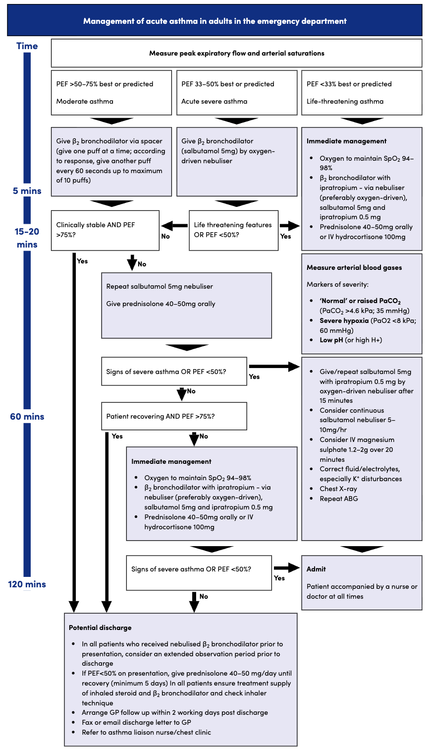 | |
Asthma algorithm. Click on image to enlarge
Escalation and critical care
Urgent senior review is required if there is:
- Failure to respond to initial therapy
- Rising PaCO₂
- Persistent hypoxaemia
- Exhaustion
- Reduced consciousness
- Silent chest
- Haemodynamic instability
Early discussion with intensive care should occur if intubation or mechanical ventilation may be required. Non-invasive ventilation (NIV) is not routinely recommended in acute asthma because evidence of benefit is limited.
Intubation should only be performed by clinicians experienced in managing the difficult airway. Positive pressure ventilation carries risks of dynamic hyperinflation, barotrauma and cardiovascular collapse.
Asthma causing cardiac arrest
Cardiac arrest due to asthma usually follows prolonged severe bronchospasm causing hypoxaemia, respiratory muscle fatigue and dynamic hyperinflation. Pulseless electrical activity (PEA) and asystole are the commonest presenting rhythms.
Potential reversible causes include:
- Severe hypoxaemia
- Dynamic hyperinflation (auto-PEEP)
- Tension pneumothorax
- Mucus plugging
- Hypovolaemia
Management should follow standard ALS principles while treating reversible causes. Important considerations include:
- Deliver high-concentration oxygen.
- Continue bronchodilator therapy where feasible.
- Administer IV corticosteroids.
- Consider IV magnesium sulfate if not already given.
- Exclude and immediately treat tension pneumothorax.
- Intubate early if required using an experienced operator.
Following intubation, ventilation should aim to minimise dynamic hyperinflation by using:
- Low respiratory rates.
- Low tidal volumes.
- Prolonged expiratory time.
- Permissive hypercapnia where appropriate.
If dynamic hyperinflation causes severe haemodynamic compromise, temporary disconnection from the ventilator for several seconds may allow trapped gas to escape and improve venous return before ventilation is recommenced.
Patients successfully resuscitated from asthma-related cardiac arrest require intensive care admission.
Disposition
Discharge may be considered when:
- Symptoms have resolved or substantially improved.
- PEF is >75% of best or predicted and maintained after treatment.
- Oxygen saturation is stable on room air.
- The patient can use inhalers correctly.
- Appropriate follow-up has been arranged.
Before discharge:
- Optimise maintenance therapy.
- Check inhaler technique.
- Provide or update a personalised asthma action plan.
- Arrange GP or asthma clinic review, usually within two working days following hospital treatment.
Admission is recommended for:
- Persistent severe symptoms.
- Incomplete response to treatment.
- Life-threatening features.
- Requirement for repeated nebulisers or oxygen.
- Significant social concerns or inability to access follow-up.
Risk factors for fatal asthma
Patients at increased risk of fatal asthma include those with:
- Previous ICU admission or mechanical ventilation for asthma.
- Previous near-fatal asthma.
- Recent hospital admission.
- Excessive reliance on short-acting β₂-agonists.
- Poor adherence to inhaled corticosteroids.
- Significant psychosocial problems.
- Food allergy with asthma.
- Poor perception of symptom severity.
Key points
- Acute asthma severity is determined by the most severe clinical feature present.
- Patients should be reassessed frequently as asthma severity can change rapidly.
- A normal or raised PaCO₂ during an acute asthma attack is an ominous sign indicating respiratory muscle fatigue.
- A silent chest represents critically reduced airflow and is a feature of life-threatening asthma.
- Oxygen should be titrated to maintain SpO₂ 94–98%.
- First-line ED treatment is oxygen (if required), repeated nebulised salbutamol, early corticosteroids and nebulised ipratropium for acute severe or life-threatening asthma.
- IV magnesium sulfate (1.2–2 g) should be considered when there is an inadequate response to initial therapy in acute severe or life-threatening asthma.
- Chest X-ray is not routinely indicated and should only be performed if an alternative diagnosis or complication is suspected.
- NIV is not routinely recommended in acute asthma.
- Early senior and critical care involvement is essential in patients with deteriorating respiratory function, exhaustion or rising PaCO₂.
- Most asthma-related cardiac arrests present with PEA or asystole and require treatment of reversible causes, particularly dynamic hyperinflation and tension pneumothorax.
Textbooks
Links


Comments
Post a Comment